How Chameleons Change Color Scientifically: Chromatophores and Crystals
For decades, classroom posters explained chameleon color with pigment cells alone: chromatophores shuffling dyes the way an artist shuffles paint. That pigment story is partly true and partly outdated. A scientific account of how chameleons change color now centers on structural color—ordered guanine nanocrystals inside iridophores whose lattice spacing can shift—while pigment chromatophores remain essential partners. The clearest modern window onto that system comes from panther chameleon research published in Nature Communications in 2015 by Teyssier and colleagues. This article walks the stack from pigments to photonic lattices, summarizes what that study changed, and marks what remains open.
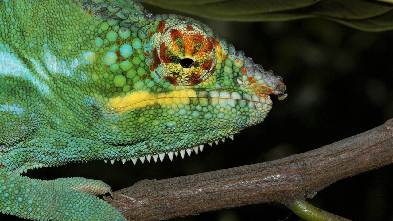
Close view of textured skin for educational context
One-sentence takeaway
Scientifically, many dramatic chameleon color changes arise when a guanine nanocrystal lattice in superficial iridophores expands or contracts under physiological control, with pigment chromatophores tinting and darkening the result—and a deeper reflective layer contributing to near-infrared heat management—while exact cellular control and species-wide generality remain active research frontiers.
That single sentence is enough to retire the paint-only cartoon without pretending every open question is closed.
From pigment story to photonic story
Pigment color is chemistry: molecules absorb some wavelengths and transmit or reflect others. Structural color is physics of arrangement: microscopic spacing and refractive-index contrasts create constructive and destructive interference, so certain wavelengths bounce back preferentially. Butterfly wings, peacock feathers, and some beetle shells are classic structural-color examples. Chameleon skin joins that club, with a twist: the structure is actively tunable on behavioral timescales.
The older pigment-only narrative struggled to explain rapid hue sweeps from blue-green toward yellow-red without inventing unrealistically fast pigment synthesis. Photonic lattices solve that puzzle: change spacing, change the reflected peak. Pigments still filter and darken; they are no longer asked to do all the work.
Speaking scientifically about chameleon color therefore means speaking in layers, interactions, and reflectance spectra—not in metaphors of living paint pots.
This shift in framing also changes what counts as evidence. A pigment-only story leans on histology of dye-bearing cells. A photonic story adds geometry, spacing statistics, and spectral curves. Both data types belong in a complete account; the 2015 panther chameleon work is famous because it bound geometry to spectra tightly enough to rewrite textbooks’ emphasis.
The chromatophore stack
Chameleon dermis organizes specialized cells in depth. Textbooks often describe a vertical stack in which different cell types occupy characteristic positions, though real tissue is three-dimensional and species can differ in detail.
Yellow and red pigment cells
Upper pigment cells commonly carry yellow (xanthophore) and red (erythrophore) pigments such as pteridines and carotenoids. These pigments absorb portions of the spectrum. Light that has already interacted with deeper reflective structures passes through these filters on the way out, so a structurally favored blue can appear green once yellow pigment sits in the optical path. Red pigments push the final appearance toward warmer tones when combined with an expanded lattice reflection.
Without these pigments, structural reflection would look different—often cooler or less saturated in the way human observers describe “chameleon colors.” Pigments are the tinting stage of the instrument.
Chemically, many yellow and red pigments in lizard chromatophores are pteridines or carotenoids (dietary carotenoids can influence reds and yellows in vertebrates generally, though species-specific details vary and should not be overstated without direct measurements). The educational point is simpler: absorbing molecules carve the spectrum after—or as—structural reflection does its selective bounce.
Melanin and darkening
Melanophores contain melanin. When melanin is dispersed into a more optically influential configuration, the skin darkens: brightness drops, colors mute, and the animal can approach brown or near-black. When melanin’s influence recedes, structural colors and yellow-red tints become more vivid.
Darkening is a major axis of change in many reptiles. In chameleons it collaborates with structural tuning: the same animal can shift hue via the lattice and shift value (lightness) via melanin. Scientifically, both axes matter for the spectra measured by instruments and for the signals other animals see.
Separating hue from lightness helps avoid confusion in popular explanations. An animal can become “darker” without becoming “redder,” or become redder without becoming darker, depending on which subsystems move. Precise language tracks which layer is active.
Iridophores and guanine nanocrystals
Iridophores are the reflective specialists. Their active ingredient in the structural story is guanine organized as nanocrystals.
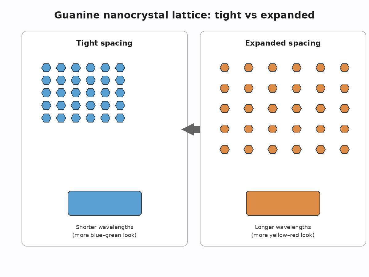
Before-and-after spacing in a nanocrystal lattice
Tight lattice vs expanded lattice
In the superficial iridophore layer of panther chameleons, guanine nanocrystals form a relatively ordered lattice. That order is what earns the comparison to a photonic crystal. When the lattice is compact, the selective reflection favors shorter wavelengths—contributing to blue-leaning appearances (modified further by overlying pigments). When physiological signals drive lattice expansion, the characteristic spacing increases and the reflectance peak moves toward longer wavelengths—yellow through red territory in display states.
The scientific punch line is geometric: hue tracking lattice constant, not a new paint job. Optical measurements and modeling in the 2015 study tied geometry to color in a way pigment redistribution alone could not.
In physical terms, periodic stacks produce wavelength-dependent interference. Increase the characteristic spacing and the wavelengths that constructively reflect tend to lengthen. Decrease spacing and the favored band shortens. Biology supplies the actuators; optics supplies the color map. That division of labor is the heart of “chameleons change color scientifically” for rapid displays.
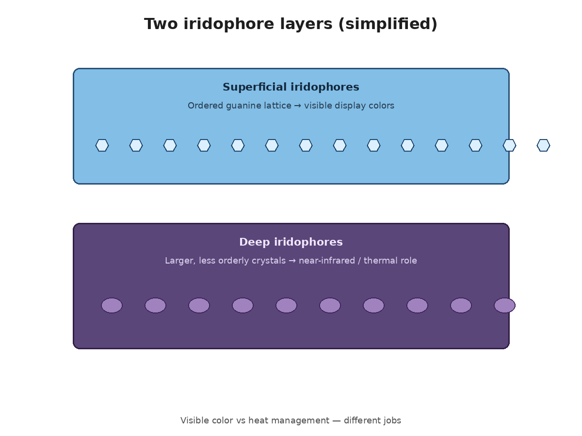
Two iridophore layers (display vs infrared reflection)
The same research distinguished two iridophore populations. The superficial population, with its ordered nanocrystals, is the display engine for rapid visible color change. A deeper population contains larger, less orderly guanine crystals and reflects strongly in the near-infrared. That deeper reflectance is discussed as a contributor to passive thermal regulation: managing solar heat load rather than painting courtship reds.
Two layers, two jobs—visible signaling optics above, heat-relevant reflection below—illustrate how “color change” in the broad sense spans both what eyes see and what thermometers would care about.
Keeping the layers distinct also prevents a common mix-up: not every reflective guanine structure in the skin is doing social billboard work. Depth and crystal organization predict function. Superficial order favors tunable visible color; deeper, coarser arrangements favor broadband near-infrared bounce in the animals studied.
What the 2015 panther chameleon research changed
Before detailed lattice measurements, structural contributions were suspected in lizards, but the panther chameleon paper made the tunable-nanocrystal mechanism concrete. Combining microscopy, spectroscopy, and physical modeling, Teyssier et al. showed that excited animals’ superficial lattices were more widely spaced than those of calm animals, matching the shift from shorter- to longer-wavelength reflectance.
That result repositioned scientific explanations:
Rapid bright displays became primarily a photonic phenomenon with pigment support.
“How chameleons change color scientifically” gained a specific object: guanine nanocrystal spacing under physiological control.
Thermal discussion gained a structural actor in the deep iridophore layer.
University summaries of the work (including coverage associated with the University of Geneva group involved in the study) helped translate the finding for broader audiences: chameleons as living photonic crystals, not merely pigment bags.
Important boundary: the study focused on panther chameleons. It transformed the template for thinking about dramatic change in that system; it did not automatically measure every chameleon species.
For science communicators, that boundary is a feature, not a bug. Landmark papers often choose a tractable, dramatic system. Later comparative work tests how far the template travels. Until those comparisons are in hand for a given species, the honest phrasing is “in panther chameleons, researchers showed…” rather than “all chameleons work exactly like this.”
Superficial display layer and deeper infrared-reflective layer
What scientists still don’t fully know
Honesty is part of scientific literacy. Several gaps remain:
Molecular and neural control of spacing. How exactly do signals inside the cell expand or compress the lattice? The 2015 paper indicated that mechanisms remained to be determined. Pathways are not fully solved in the public literature that educators should lean on; inventing a neat “ion X does Y” story would overclaim.
Camouflage versus signaling weights. How much background matching contributes relative to social and thermal drivers differs by species and context. Absolute slogans either way mislead.
Species generality. Panther chameleons are not stand-ins for all chameleons. Comparative work is needed before assuming identical lattices, speeds, or functional rankings elsewhere.
Development and plasticity. How lattices are built as animals grow, and how nutrition or health affect photonic performance, are richer questions than a single landmark paper can close.
Receiver-side vision. Signals evolve relative to what receivers can see. Birds, snakes, and other lizards differ in visual systems; mapping chameleon spectra onto those systems is ongoing ecological work, not a finished table in every textbook.
Marking these gaps keeps educational writing aligned with evidence rather than with tidy myths. Students can learn the photonic mechanism and learn that science remains open—those lessons reinforce each other.
Glossary
Chromatophore — A pigment-containing cell (for example, yellow/red pigment cells or melanophores) that influences color by absorbing light.
Iridophore — A reflective cell containing guanine crystals; in chameleons, superficial iridophores enable tunable structural color, while deeper ones can reflect near-infrared.
Structural color — Color produced by microscopic physical structure (interference, diffraction, scattering) rather than by pigment absorption alone.
Photonic crystal — A material with periodic refractive-index structure that affects which wavelengths propagate or reflect; biological lattices of guanine nanocrystals can act in this way.
Guanine nanocrystal — Nanoscale crystal of guanine arranged in iridophores; spacing of the array tunes reflectance.
Melanin — Dark pigment in melanophores that reduces brightness and can mute other colors when optically influential.
Near-infrared (near-IR) — Light just beyond the visible red; relevant to heat and to deep-iridophore reflectance in studied chameleons.